Mild surface dryness
Observed: Light, scattered surface flaking without marked redness.
What this can indicate: The scalp barrier may benefit from gentler cleansing and hydration.
Your Scalp Scan Report
The scan shows generally healthy follicular grouping with mild dryness and some variation in hair-shaft thickness near the crown. These are useful areas to support and compare at a future rescan.
Follicular grouping is preserved across assessed zones. Mild surface scaling and moderate shaft-diameter variability are most visible at the vertex; no urgent inflammatory signs are represented in this sample.
Most visible units contain two or more hairs.
No significant redness is represented in the assessed zones.
Practical actions based on this scan — start here after reading the summary.
Hair density and thickness in each zone scanned. Estimates are visual, from trichoscopy footage.
| Area | Density | Hair thickness | What it means |
|---|---|---|---|
| Crown | Within expected range |
Mild
~18% finer strands
|
Findings in this zone, with a mild mix of finer and thicker hairs, which is often stable.
|
| Mid-scalp | Within expected range |
Minimal
~11% finer strands
|
Findings in this zone, with mostly uniform hair shaft thickness.
|
| Frontal | Within expected range |
Minimal
~13% finer strands
|
Findings in this zone, with mostly uniform hair shaft thickness.
|
Finer-strand percentages are visual estimates from scan footage — a guide, not a follicle count.
Observations that differ from a healthy baseline and are worth tracking over time.
Key frames selected from your scan where each finding is most clearly visible.
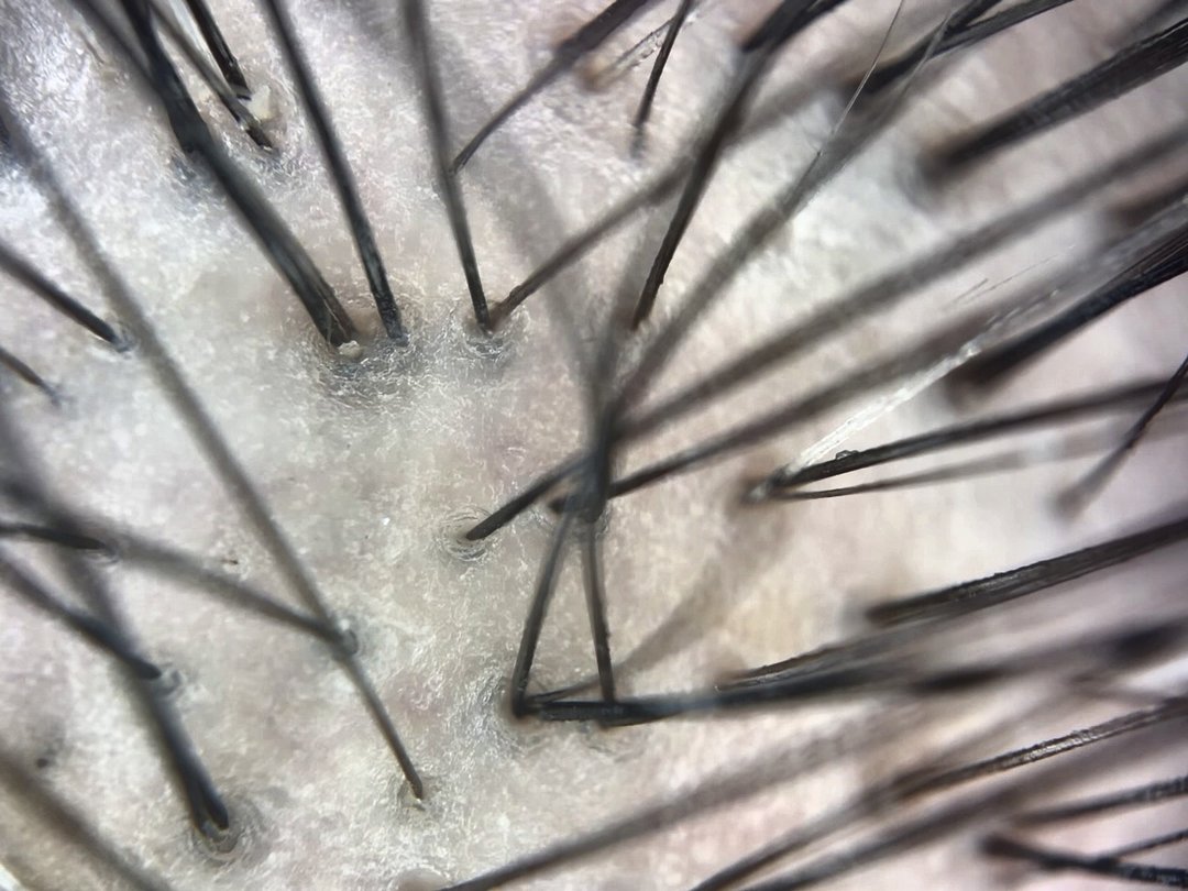
Several follicles show more than one hair shaft, creating a useful baseline for future scans.
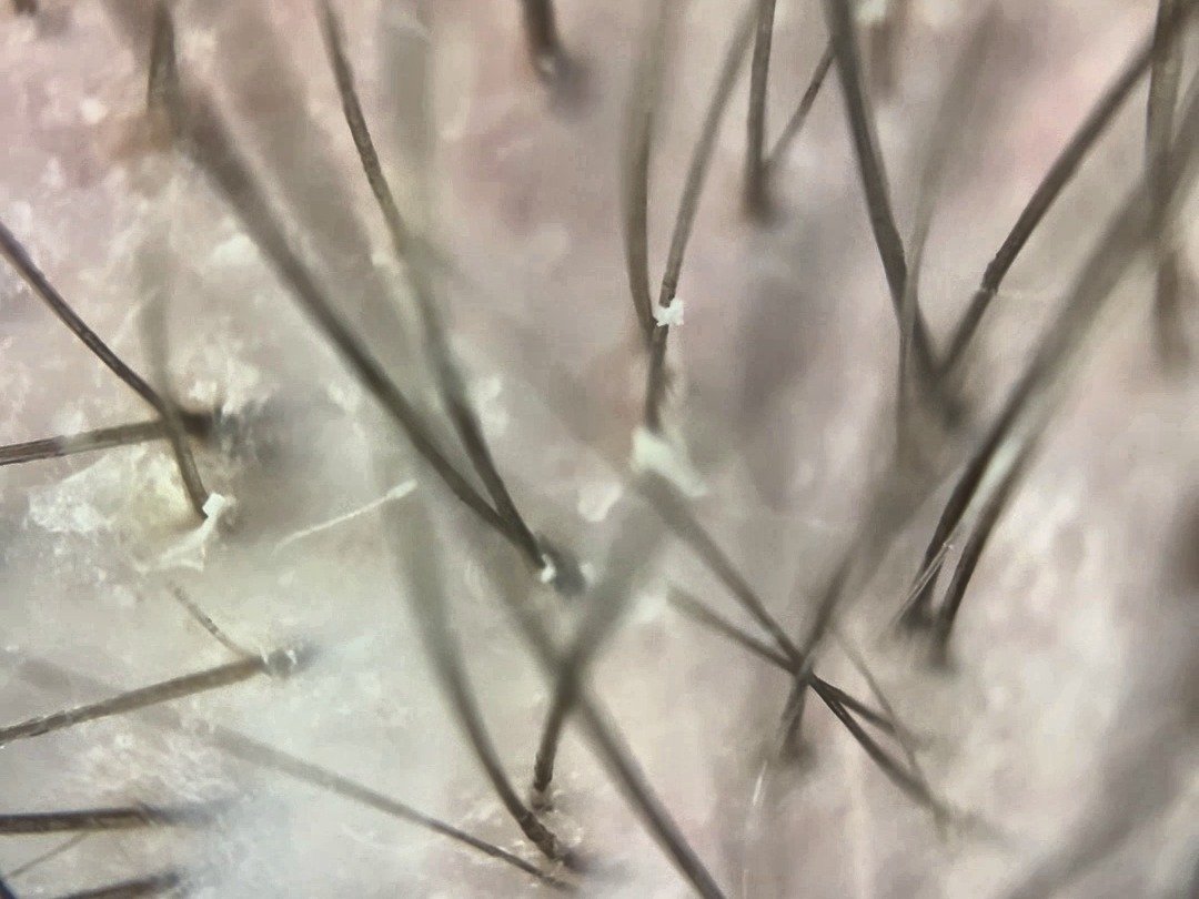
Small, light flakes are visible near some hair bases without marked redness.
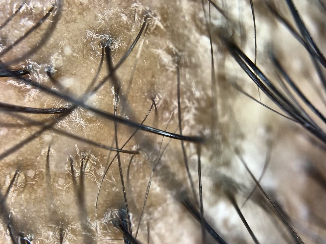
A mix of finer and thicker shafts is visible, providing a clear area to compare at the next scan.
Five domains reviewed in every scan. Status and observations — see Findings above for what to watch.
Consistent crown, mid-scalp, and frontal views make follow-up conversations more useful than unrelated photos.
HumanTouch reviewed close-up scalp video from your visit and selected the clearest frames for this report.